
 Medically Reviewed
Written by Pooja Rawat, Medical Content Writer
Posted On: 27 Oct 2026,
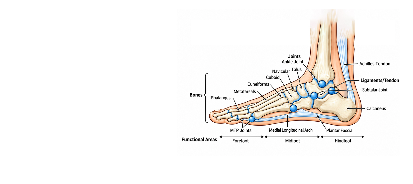
Have you ever wondered how many bones your foot has? The answer is 26. Your ankle, however, has two more bones: the tibia and the fibula. So together, the foot and the ankle have 28 bones, which are categorised into three groups. The hindfoot (back of the foot), midfoot (middle of the foot), and forefoot (front of the foot). To hold these bones in place, we have 33 different joints along with strong ligaments, tendons, and muscles. All these work together to help us perform normal activities today, like standing, walking, running, jumping, etc
It is quite easy to forget how much work your feet do throughout the day until they start to hurt. You hold your feet and gently press them, and at that point, the only thing you want to know is what exactly lies beneath that ache and what is causing it: a bone, a tendon, a ligament, or something else.
This blog will help you understand all about foot and ankle anatomy. From the number of bones and joints to the ligaments and muscles, this blog covers everything you need to know about your feet. You will also get to know about some common problems related to every part of the feet to help you understand them better, so that the next time your feet are hurting, you know exactly what is going wrong.
Read Also: Ankle Injuries Treatment in Dwarka: Best Doctors & Clinics Near You
According to the chapter Anatomy, Bony Pelvis and Lower Limb: Foot and Ankle published by StatPearls, our foot is made of 26 bones, out of which 7 are tarsal bones, 5 are metatarsals, and the remaining 14 are phalanges (toe bones).
| Area of the foot | Main bones | What it does |
| Hindfoot | Talus, calcaneus (heel bone) | Forms the ankle and heel, takes the first load when you step |
| Midfoot | Navicular, cuboid, three cuneiforms | Forms the arch, absorbs shock |
| Forefoot | Five metatarsals, 14 phalanges | Pushes off the ground, helps balance |
All these bones are surrounded by joints, ligaments, tendons, muscles, nerves, and blood vessels; all have an important purpose and role to play. These work like a team to carry your body weight and allow you to walk, run, climb and balance.
Read Also: Sprained Ankle Explained: Types, Symptoms, and Treatment Options
If we have to define the ankle in simple words, it is the point where the leg ends and the foot begins.
The ankle joint consists of three bones: the tibia (the shin bone), the fibula, and the talus. The tibia and fibula together make a socket in which the talus fits like a hinge. It is this hinge that allows your foot to move up and down.
There are two bumps that you can see and feel very easily. The first one is the inner bump, or the medial malleolus, which is formed by the tibia. The second one is the outer bump, or the lateral malleolus. This is formed by the fibula.
Surprisingly, there is a third bump too, but it cannot be seen or felt easily. This lies at the back of the tibia.
Read Also: Difference Between Ankle Sprain And Ankle Strain?
Do you know how many bones are there in your foot and ankle? The answer is 26, and including the lower ends of the tibia and fibula, there are 28.
Anatomically, the foot is divided into three sections. These are:
The foot and the ankle together have around 33 joints between them. Here are some that are worth knowing.
These are strong tissues with a rope-like structure that help to connect the bones and keep the joints steady. The plantar fascia is a thick band of connective tissue found in the foot. It connects the calcaneus, or heel bone, to the forefoot.
Ligaments found on the lateral, outer side of the ankle are known as lateral ligaments. These include the anterior talofibular ligament and the calcaneofibular ligament.
On the inner side of the ankle is the thick deltoid ligament, and above the ankle are the syndesmotic ligaments, which are responsible for holding the tibia and fibula together.
The muscles are soft tissues that are elastic in nature. They contract and relax to cause movement. The human body contains 650 muscles, and out of these, 20 are found in the foot. These are categorised into two categories: intrinsic muscles and extrinsic muscles. The intrinsic muscles are present within the foot and are responsible for the movement of the toes. The extrinsic muscles are located outside the foot, usually towards the lower leg. Your calf muscle is also an extrinsic muscle that helps in moving your feet.
A tendon is like a dense band of fibrous connective tissue that connects the muscles to the bones. Tendons found inside the foot include”
Our feet aren’t flat, even though they look flat when we stand. Each foot has three arches. The medial longitudinal arch runs along the inner side and is the highest of the three. The lateral longitudinal arch runs along the outer side. The transverse arch curves across the middle of the foot.
These three arches help to absorb shock when we walk. The shape can vary from one person to another, and while some have a departure, others have a very mild one. People with the latter are said to have a flat foot
The sole of the foot appears very thick and strong because it has been made for taking pressure and withstanding friction. Under it is a thick pad of fat, which is thickest at the heel. This acts as a cushion. Inside this layer are the plantar fascia and small muscles of the foot, which are arranged in four different layers, below which lie the bones.
The main nerve connected to the sole of the foot is the tibial nerve, which divides into the medial and lateral plantar nerves. It passes behind the inner ankle bone through a narrow passage. This is known as the tarsal tunnel.
The arteries responsible for supplying blood to the foot are known as the posterior tibial artery and dorsalis pedis artery. In people with diabetes and similar conditions, the nerves can get damaged, restricting the flow of blood to the foot. This is the reason why patients with diabetes are recommended to go for regular foot checks.
The foot and the ankle may seem like simple structures, but they are very complex from within, and so is the mechanism. These are made up of bones, joints, ligaments, tendons, muscles, and arches all working together, like a team to ensure smooth movement. No matter how insignificant they may seem, taking care of your feet and ankles is very important to ensure pain-free movement If you want to know more about your foot and ankle or if you're experiencing any symptoms related to these, you can consult Dr Ishwar Bohra for expert care.
Disclaimer: This blog is for general information only. It is not a substitute for medical advice, diagnosis or treatment. Please consult a qualified doctor about your symptoms.
References
Q1. What are the main parts of the foot?
A: The foot is divided into three main parts: the hind foot, the midfoot and the forefoot. These are joined by joints, ligaments, tendons, muscles, nerves, and skin.
Q2. How many bones are in the foot and ankle?
A: There are 28 bones in the foot and ankle. 26 of these are in the foot, whereas two are connected to the ankle joint
Q3. What are the bones of the foot called?
A: The bones of the foot are called the talus, calcaneus, navicular, cuboid, cuneiforms, metatarsals and phalanges
Q4. What is the largest bone in the foot?
A: The calcaneus, or heel bone.
Q5. What is the ventral aspect of the foot?
A: The ventral aspect of the foot refers to the underside of the foot, which is also known as the plantar surface or sole.

Staying abreast with the latest information is the best way to combat diseases in early stages and live a healthy life. Read the latest news & updates here to learn about the recent advancements in joint care and therapeutics.